Is Your Child’s Skin ‘Peeling’ Between Their Toes?
Tinea Pedis or ‘Athlete’s Foot’
According to Spain and Espinoza, 2019: About 20% of the entire population worldwide suffers from some type of mycosis, of which more than 70% occur in the most vulnerable people, namelychildren and adolescents.. The aetiological agents vary depending on the climate, cultural and socio-economic characteristics of the population. (1) (1)
Infections of the skin, hair and nails caused by dermatophytesare calledtinea (ringworm). They are the most common superficial mycoses worldwide. According to studies by authors Jimenez et al (2017), tinea is classified according to the body region affected,for example, tinea corporis involves arms, trunk and legs; tinea capitis involves the scalp, and tinea pedis on the feet. (2) (2)
What is Tinea Pedis or ‘Athlete’s Foot’?
Tinea pedisis the condition commonly known as ‘athlete’s foot’ and is usually caused by Trichophyton rubrum, Trichophyton mentagrophytes interdigitale or Epidermophyton floccosum, and more than one fungus may be present. These infections are considered a public health problem due to their contagiousness and recurrent nature. (3). (3)
Why does it develop?
The geographic distribution of this pathology is worldwide and it is undoubtedly the most common dermatophytosis. It occurs mainly in the summer monthsbut may persist inactive during thewinter.. The incidence is high in tropical regions and is higher in people who wear thick socks and very closed shoes and in peoplewith hyperhidrosis, as well asfrom the use of public swimming pools, showers, towels or the use of other people’s socks and shoes.. There is currently a high incidence in schoolchildren due to the constant use of occlusive footwear(4).
Theclinical picturemay vary, but three forms of tinea capitis can be distinguished in which the most frequent symptoms are pruritus and burning: (4)
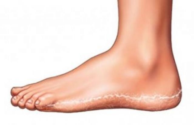
- – Intertriginous form located in the interdigital grooves and in the crease of the plantar aspect of the toes. These lesions consist of maceration and/or fissures and are characterised by pruritus as the main sign associated with malodour.
- – Vesicular form located on the plantar surface of the foot. It is characterised by numerous vesicles of viscous content that tend to rupture and leave erosions and crusts associated with pruritus.
- – Hyperkeratotic form is considered as a chronic variety. It consists of hyperkeratotic plaques that flake off abundantly.
How is it identified?
In order to identify them, it is necessary tocarry out a good anamnesis, an exhaustive physical examination and performcomplementary tests such as live examination by optical microscopy or culture, with prior sampling either by scraping or by milling. In this way we will achieve a definitive diagnosis in this type of infection. (5-8) (5-8)
A correct differential diagnosis with pathologies such as interdigital candidiasis impetigo, infection of the interdigital space, by pseudomona aeruginosa erythrasma and, keratolysispunctata must be established (5-6)..(5-6)
The most important prerequisite for initiating drug treatmentis a positive result on culture ordirect microscopic examination.(9-11)
What is your treatment?
The antifungal treatment used may be topical or oral, in both cases failuremay be due not only to drug or pathogen resistance, but also to incorrect application of the treatment or cessationof treatmentbefore the prescribed regimen.
It is thereforeessential that the patient is fully informed and understands the need to properly follow the professional’s instructions in order to achieve treatment success.. In addition, it is crucial to reduce risk factors as much as possible and to complement treatment with preventive measures.. Preventive guidelines that every patient should take into account when applying treatment include: (5-7) (12-16)
- – Proper cleaning and drying of the affected areas.
- – Do not walk barefoot in public toilets and showers.
- Wearing non-enclosed footwear and absorbent socks, avoiding occlusive or poorly breathable clothing
- Use antifungal powders for footwear and/or socksif they are prone to recurrence.
- Inform the patientabout the importance of not interrupting treatment even if symptoms subside a few days after starting treatment.
- – Avoid applying large amounts of cream to affectedinterdigital spaces to prevent maceration.
-
España, S., & Espinoza, T. (8 de Marzo de 2019). Situación de la Micosis Superficial en Ecuador. Trabajo de Titulación. Guayaquil, Guayas, Ecuador: UCSG. Obtenido de http://repositorio.ucsg.edu.ec/bitstream/3317/12568/1/T-UCSG-PRE-MED-ENF524.pdf
-
Jiménez, H., Briseño, G., Vásquez, E., & Arenas, R. (julio de 2017). Tinea pedis y otras infecciones podales: datos clínicos y microbiológicos en 140 casos. Dermatología Cosmética, Médica y Quirúrgica, 15(3), 156-161. Obtenido de https://www.medigraphic.com/pdfs/cosmetica/dcm-2017/dcm173c.pdf
-
Sabogal, M., Jiménez, H., Morales, C., Alvarado, Z., & Colmenares, C. (2019). Micosis en los pies: descripción clínico-epidemiológica en un centro de referencia de Bogotá, Colombia. Infectio, 39-34. Obtenido de http://www.scielo.org.co/pdf/inf/v23n1/0123-9392-inf-23-01-00039.pdf
-
Blanco B et al. Temas de dermarología pediátrica. Pautas diagnósticas y terapéuticas.Ateproca.2005.
-
Peña, A. Atlas de dermatología del pie. Madrid: Médica Panamericana; 2007
-
Zalacain, A. Ogalla, JM. García-Patos, V. Atlas y sinopsis de enfermedades cutáneas del pie. Barcelona: Edika Med; 2008
-
Garcia, FJ. Guia clínica para el tratamiento de las micosis del pie. Madrid: Fco. Javier García; 2003
-
Kumar, V. Tilak, R. Prakash, P. Nigam, C. Gupta, R. Tinea Pedis- An update. Asian Journal Of Medical Sciences 2 (2011) 134-138.
-
Chauvin, M. Viguie, C. Kienzier, JL. Larnier, C. Novel, single-dose, topical treatment of tinea pedis using terbinafine: results of a dose-finding clinical trial. Mycoses. 2008 Jan;51(1):1-6
-
Ortonne, JP. Korting, H. Viguie, C. Larrier, C. Savaluny, E. Efficacy and safety of a new single-dose terbinafine 1% formulation in patients with tinea pedis (athlete’s foot): a randomized, double-blind, placebo-controlled study. J Eur Acad Dermatol Venereol. 2006;20:1307–13
-
Li, R. Xu, J. Xi, L. Fu, M. Zhu, M. Efficacy and Safety of 1 % Terbinafine Film-Forming Solution in Chinese Patients with Tinea Pedis: A Randomized, Double-Blind, Placebo-Controlled, Multicenter, ParallelGroup Study. Clin Drug Investig. 2014;34(3):223–30
-
Nenoff P, Krüger C, Schaller J, Ginter-Hanselmayer G, Schulte-Beerbühl R, Tietz H-J. Mycology – an update Part 2: Dermatomycoses: Clinical picture and diagnostics. JDDG J der Dtsch Dermatologischen Gesellschaft [Internet]. 2014;12(9):749–77
-
Puig, L. Vilarrasa, E. Tiña interdigital de los pies (pie de atleta): su diagnóstico y últimos avances en su tratamiento. Madrid: sanidad y ediciones; 2008
-
Weinstein A, Berman B. Topical treatment of common superficial tinea infections. American Family Physician 2002;65(10):2095-2102
-
Gupta AK, Chow M, Daniel CR, Aly R. Treatments of tinea pedis. Dermatologic Clinics. 2003; 431–62
-
Conejo, A., Martínez, M., & Alfayate, S. (21 de 11 de 2018). Dermatofitosis o tiñas. Obtenido de Guía ABE: https://guia-abe.es/temas-clinicos-micosis-cutaneas

