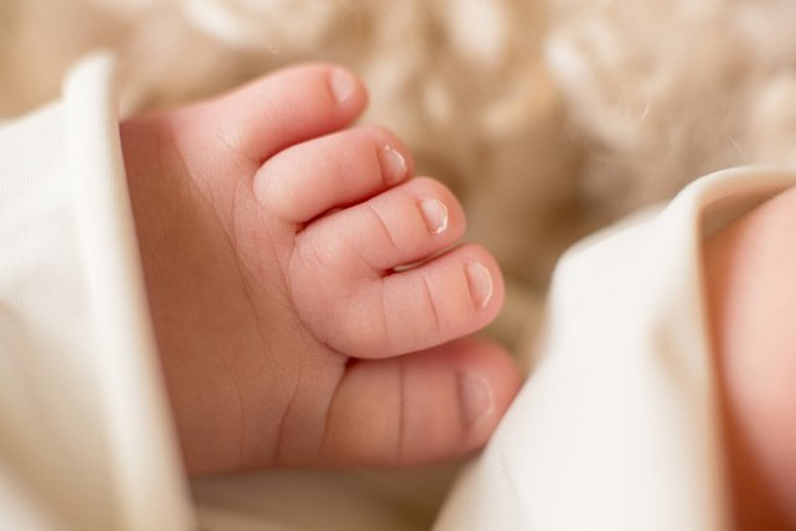
SYNDACTYLY IN CHILDREN
APERT SYNDROME
Congenital pathologies of the forefoot can be classified as follows malformations (during embryonic development) and on the other hand, deformities produced in the foetal produced in a foetal period when the foot is already configured in a normal way. The latter are easier to heal. Syndactylybelongs to the group of malformations, where anatomical defects occur (1).
What is syndactyly?
Syndactyly is a congenital malformation of bilateral genetic origin or it can occur sporadically. or it may occur sporadically. It occurs in both hands and feet, so that they are It occurs in both hands and feet, so that the soft (simple) parts of the feet and hands are joined together, or there may even be cases of bony union (bony union). cases of bony union (complex). All this is due to a failure in embryonic development. Its prevalence is 3-10 cases per 10,000 births, mostly in males (2,3).
 What does it affect?
Finger involvement is variable. In terms of treatment, there are several options: surgicalto avoid impingement of the longest toe due to footwear, or in the other footwear, or in the other spaces if there are no functional abnormalities, an observational treatment is chosen. observational treatment. The children’s families are informed of the risks, especially in terms of scarring (keloids) in the event of surgical treatment (4).
How is it treated?
Several techniques are used for simple and complex congenital syndactyly, in order to free up the necessary space between the affected fingers and thus allow independent functionality (5).
Some syndromes associated with this pathology are ectrodactyly syndrome, ectodermal dysplasia and ectodermal dysplasia, ectodermal dysplasia and cleft lip/palate. It affects the handsbut syndactyly also occurs syndactylyalso occurs in the toes (third and fourth; fourth and fifth toes) (6,7,8).
Other scientific studies have demonstrated its association with the syndrome of Poland syndrome, which occurs in this case in the fingers. It is the most frequent of all syndromes that present syndactyly.
On the other hand, we are presented with cases of simple incomplete syndactyly which is This may be due to a structural alteration of the forefoot, such as the first metatarsal. such as the case of the short first metatarsal, showing a false image of syndactyly (9,10,11).
What is Apert’s syndrome?
Apert’s syndrome is characterised by premature closure of the cranial sutures, which results in sutures, resulting in consequent malformations of the face, hands and feet. This conditionis of concern in such a way that syndactylies appear. It is of congenital congenital and is considered a rare disease
This pathology can appear bilaterally and symmetrically, thus associating bony syndactyly with membranous syndactyly (14,15).
When should it be diagnosed?
The diagnosis of this type of pathology should be made as early as possible in order to be able to act and rapid action can then be taken to initiate treatment. Astrong emphasis is placed on prevention, as we know that the disease is preceded by very advanced paternal age (15,16).
One of the repercussions of a late diagnosis may be the risk of airway compromise, hearingand eye movements or evenIQ compromise. Early surgeries are therefore recommended for age ranges and stages of the disease are therefore recommended. In the case of syndactyly of the feet orthopaedic and plastic treatments are offered in order to relieve the soft tissue involvement, together with the soft tissue involvement, together with the possibility of performing toe clamping with the “thumb pinching” (17,18,19).
Bibliography
-
Rampal V, Giuliano F. Forefoot malformations, deformities and other congenital defects in children. Orthop Traumatol Surg Res [Internet]. 2020;106(1):S115-
23. Disponible en: https://doi.org/10.1016/j.otsr.2019.03.021 -
Jordan D, Hindocha S, Dhital M, Saleh M, Khan W. The epidemiology, genetics and future management of syndactyly. Open Orthop J. 2012;6:14–27.
-
K. McGarry, S.Martin, M.McBride, W.Beswick, H.Lewis. The Operative Incidence of Syndactyly in Northern Ireland. A 10-Year Review. Ulster Med J. 2021 Jan; 90(1): 3-6.
-
Jouve JL, Jacquemier M, Casanova D, Launay F, Bardot J, Magalon G, Bollini
G. Anomalies congénitales des Orteils (Métatarsus adductus et hallux val- gus exceptés) Monographie du Groupe D’Etude en Orthopédie Pédiatrique. Sauramps Médical 2001:187–201. -
Mende K, Watson A, Stewart DA. Surgical Treatment and Outcomes of
Syndactyly: A Systematic Review. J hand Surg Asian-Pacific Vol. 2020;25(1):1-12 -
Meza Escobar LE, Isaza C, Pachajoa H. Ectrodactyly, ectodermal dysplasia and cleft lip/palate syndrome, report of a case with variable expressivity. Arch Argent Pediatr 110(5):e95-8, 2012
-
Celik TH, Buyukcam A, Simsek-Kiper PO, Utine GE, Ersoy-Evans S, Korkmaz A, et al. A newborn with overlapping features of AEC and EEC syndromes. Am J Med Genet A 155A(12):3100-3, 2011
-
Marwaha M, Nanda KD. Ectrodactyly, ectodermal dysplasia, cleft lip, and palate (EEC syndrome). Contemp Clin Dent 3(2):205-8, 2012
-
Malik S. Syndactyly: phenotypes, genetics and current classification. Eur J HumGenet. 2012; 20: 817-824
-
Bosse K, Betz RC, Lee Y, Wienker TF, Reis A, et al. Localization of a gene for syndactyly type 1 to chromosome 2q34-q36. Medscape [Sede web]. New York: Deune E; 2013 [Consulta 2 de febrero de 2018). Syndactyly. Disponible en: http://emedicine.medscape.com/article/124
-
Jordan D, Hindocha S, Dhital M, Saleh M, Khan W. The Epidemiology, genetics and future Mangement of Syndactyly. Open Orthop J. 2012; (6):14-27
-
Álvarez A, Zaldivar del Campo F, Pérez LA. Acrocéfalo-sindactilia tipo I: Síndrome de Apert: presentación de un caso. Rev cienc méd habana; 13(1), ene- jun 2007
-
Carro Puig E y Fenandez Braojos L. Sindrome de Apert. Presentación de un
caso. Rev Cubana Pediatr 2005; 77 (3-4) Acceso: 25 marzo 2006. Disponible en: http://scielo.sld.cu/scielo.php?script=sci_arttext&pid=S00347531200500030000 9&lng=es&nrm=iso&tlng=es -
Habenicht R. Transverse soft tissue dis- traction preceding separation of com- plex syndactylies. J Hand Surg 2015;1- 7
-
Barro M, Ouedraogo YS, Nacro FS, Sanogo B, Kombasséré SO, Ouermi AS, et al. Apert syndrome: Diagnostic and management problems in a resource- limited country. Pediatr Rep. 2019;11(4):78-80
-
Torres Salinas CH, Lozano Ccanto B, Damián Mucha M. Síndrome de Apert. Repercusiones de un diagnóstico y abordaje tardío. Pediatria (Santiago). 2021;53(4):153-7.
-
Poggiani C, Zambelloni C, Auriemma A, Colombo A. Acrocephalosyndactyly, Apert type, in a newborn: Cerebral sonography. J Ultrasound. 2007;10(3):139– 42.
-
Wenger TL, Hing A V, Evans KN. Apert Syndrome Summary Genetic counseling. 2022;1-26.
-
Stauffer A, Farr S. Is the Apert foot an overlooked aspect of this rare genetic disease? Clinical findings and treatment options for foot deformities in Apert syndrome. BMC Musculoskelet Disord. 2020;21(1):1-7


